Isthmocele and secondary infertility
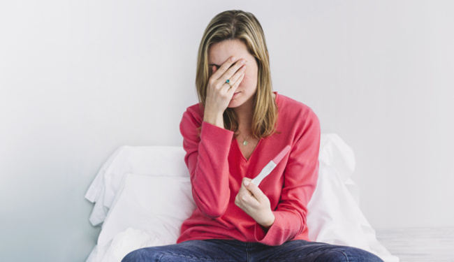
In assisted reproduction we talk about secondary infertility when a couple has already had one child but is now struggling to conceive a second child after a year of unprotected intercourse. There may be many reasons why a couple is suffering from secondary infertility. Specifically, one of the causes we see in fertility clinics are scar defects after caesarean section delivery. This is known as an isthmocele.
Taking current data into consideration, the increased number of caesarean section deliveries has also demonstrated an increased incidence and diagnosis of isthmoceles. Today we want to talk to you about this issue, why it can occur and what symptoms and diagnosis it involves.
What is an isthmocele?
An isthmocele is a partial dehiscence, or what we commonly refer to as a scar defect, which is produced following the process of a hysterotomy (the surgical opening of the uterus during a caesarean section), which leaves a sacculation or pouch‑like cavity at the site of the hysterorrhaphy, or surgical suturing, of the incision site.
How and why is an isthmocele caused?
In order for an isthmocele to be produced, the woman first needs to have undergone a caesarean section delivery (surgical procedure). While it’s true that the mechanism which causes this complication is unknown, its incidence is related to the number of prior caesarean section deliveries.
Some recent studies have estimated that an isthmocele could appear after the first C‑section delivery 60% of the time, and after three C‑sections this incidence could reach 100%.
In addition, the appearance of this complication is also related to the surgical technique used, the material and the type of suturing. The difference in size between the anterior and posterior edge of the surgical incision could also play a role, as well as the position of the woman’s uterus, the most frequent position being retroflexed.
What are the symptoms of an isthmocele?
The truth is that not all isthmoceles present clinical symptoms. At any rate, the most common symptoms include post‑menstrual bleeding, dyspareunia (pain during intercourse) and abdominal pain.
Isthmoceles have also been linked to sterility and secondary infertility due to difficulties for the spermatozoids to penetrate the uterus, as well as the detrimental effects on sperm that can occur in the cervical canal.
An isthmocele may also have a negative impact on implantation if the pouch‑like cavity contains products of menstruation that can later leak out and enter into the endometrial cavity, possibly lowering the likelihood of embryo implantation.
How is an isthmocele diagnosed?
The diagnostic process for an isthmocele is clinical and requires the help of imaging techniques: an endovaginal ultrasound and a hysteroscopy.
During the endovaginal ultrasound the size of the defect is measured: it is observed as an ellipsoidal or triangular eco negative image, so both diameters can be measured and its area can then be calculated.
The diagnostic hysteroscopy provides direct evidence of the defect, determines its severity and makes it possible to decide whether it should be surgically managed with a hysteroscopy or a laparoscopy.
On occasion, magnetic‑nuclear resonance imaging has been used to decide which surgical approach should be applied to correct the defect depending on the size, in millimetres, of the thickness of the anterior surgical edge.
How is an isthmocele treated?
Treatment of an isthmocele requires surgery only in cases presenting clinical symptomatology or if the patient suffers from infertility and there is no other known cause.
The treatment performed is a resectoscopic correction by hysteroscopy with the intention of reducing the size of the pouch‑like cavity defect, in addition to coagulating and closing the site of the surgical incision. When it is a wide, deep defect that cannot be treated by hysteroscopy, surgery must be performed by laparoscopy in order to suture the pouch‑like cavity.
There are still very few published cases on this technique, but the initial results are promising. In fact, women have been able to get pregnant naturally in the majority of cases just a few months after the surgical correction. Patients, however, are advised to wait about three months before trying to conceive again if a hysteroscopy was performed, and six months if a laparoscopy was performed.